Charcot Foot Reconstruction part 2
Charcot Foot Reconstruction part 2
In a previous post you were introduced to the concept of the charcot foot and its collapse. In This post I will talk about the innovative ways we correct this foot and save these legs from being amputated.
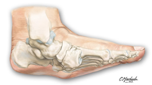
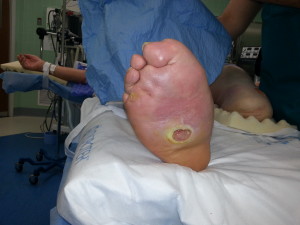
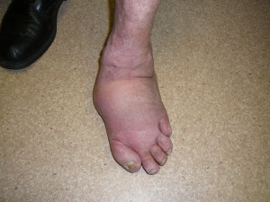
Charcot foot reconstruction has gone through an evolutionary process from amputation to placing the bones in the corrected areas to fusing the diseased joints. There has been an evolution of the hardware used from screws to plates to locking plates and to beaming the columns of the foot. The downside to single stage corrections also known as acute corrections is that one gets a smaller foot because allot of bone needs to be resected out to obtain the correction then the screws and plates are put in to maintain the correction. During that acute correction there is also a risk of pulling on arteries and nerves beyond its ability to compensate in one setting.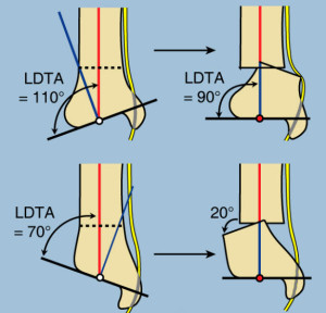
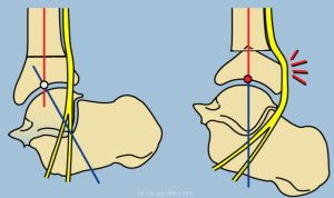 As you can see from the pictures on the left when you move the bones in the foot around you can pull on the nerves of the foot. In a charcot foot which is usually in a rockerbottom position you can stretch and pull on the posterior tibial nerve and the posterior tibial artery that runs along with it. As well the dorsalis pedis artery and the dorsal foot nerves are at risk. Historically many of these procedures have failed in the past because the surgeon attempted to do the correction in one shot and thereby putting the nerves and arteries of the foot at risk. In upcoming blogs we will discuss how to perform the 2 stage procedure ( do not do this at home) and you will see some x rays of the final product.
As always feel free to email me with any questions.
