Category: Uncategorized

  • Ankle Dislocation Robert Griffin RG III

    Ankle Dislocation Robert Griffin RG III

    ankle dislocation
    ankle dislocation
    ankle dislocation
    ankle dislocation
    ankle dislocation
    ankle dislocation fixed with Taylor spatial frame

    So by now you heard about Robert Griffin the third and his ankle dislocation. What does that mean and what are its implications. After watching the video carefully the mechanism of injury is one that the foot everts from under the leg. Another way to say it is that the leg and the foot instead of aligning straight the foot was forced into an extreme position away from the leg. This is less common than the usual ankle sprain where the foot rolls in and under the leg, his rolled out and away from the leg. The main concern when this happens it an extreme pull on the posterior tibial nerve and artery, that can cause severe nerve damage and loss of feeling in the foot. In this case there was no associated fracture but sometimes that is not the case. Even if there is no fracture the ligaments are probably torn. Also another concern is the posterior tibial tendon can get caught under the medial malleolus of the ankle. That can require surgery to put back in the proper position. The ligaments that we are concerned with are the deltoid ligament complex and it is important to evaluate the syndesmotic ligament of the ankle and sometimes the spring ligament of the foot. An MRI is best for evaluating all those structures. If the foot is going numb or there is a loss of color to the foot or toes ( they are turning white) that is a surgical emergency and the dislocation must be immediately reduced.

    The big concern is what is the recovery. How quickly can one get back to playing professional football. The answer lies in how extensive was the damage and if surgery is necessary. if surgery is necessary his season is over. If not he may be able to make it back if the Redskins make it to the playoffs. It will be interesting to see over the next few days when more extensive reports about his MRI are released.


  • Plantar plate tears part 2

    Plantar plate tears part 2/ In the last segment we discussed what is the plantar plate and what plantar plate tears are.  In this post I will discuss how are Plantar plate tears diagnosed and get into the treatment options.

    Many people will come in with pain in the balls of their feet and the doctor will quickly diagnose a mortons neuroma. The error in that is that there are many other diagnosis besides that which have a totally different treatment. The exam done is called a modified Lachmans test where i can feel if the second metatarsal phalangeal joint 9 which is by the ball of the foot under the second toe if there is a instability or mild subluxation i would go on to do an x ray.

    Plantar plate dislocation
    Plantar plate dislocation
    Plantar plate dislocation
    Plantar plate dislocation

    The x ray will NOT be able to diagnose a plantar plate tear . However it will be able to rule out a stress fracture of the second metatarsal. Also it can give us an insight if one has a long second metatarsal which can us an insight into the foots biomechaniccs and can be a precursor to a plantar plate tear. This is what differentiates going to a shoe or running store and getting a pair of orthotics off the shelf , they can not do an x ray to see whats happening. Once the x ray is negative for stress fractures that is where the next step comes in . I order an MRI arthrogram. The procedure that is performed is a common procedure for hip and shoulder capsules. A dye is injectes into the joint , if the dye leaks out the capsule is torn- in our case the capsule of the second metatarsal phalangeal joint (MPJ).

    Notice the dye leaking out of the joint
    Notice the dye leaking out of the joint
    Notice the dye leaking out of the joint
    Notice the dye leaking out of the joint

    as you can see clearly in these pictures the only clear way to diagnose thes injuries is with this exam.

    Here at the Advanced Foot and Ankle Center of San Diego we have years of experience diagnosing and treating these issues.

    In the next post we will discuss the various treatment options and how to get you back out running as soon as possible.MRI showing Plantar plate tear

    MRI showing Plantar plate tear
    plantar plate
    MRI showing Plantar plate tear
  • Lateral Ankle Instability part one

    Lateral ankle instability can be a debilitating condition.What is it? How does it happen? Is it common? It can happen many ways. Women seem to be more prone to getting it than men. Why is that? Women have ligaments that are more lax- they have a hormone called relaxin that helps the pelvis expand during pregnancy and child birth. So their ankles  or other joints can be affected as well.  Basically lateral ankle instability is what it says, you have an unstable ankle joint regarding the outside (lateral) ligaments. Symptoms include multiple sprains, a feeling of instability constant pain and swelling. It can be torn ligaments to torn peroneal tendons and sometimes the ankle joint can have an osteochondral defect from the multiple sprains. It can make exercise and even daily walking painful. Many times it starts with a sprain that does not heal properly. I have seen many misdiagnosed ankle ligament tears that were thought to be sprains. MRI was not taken and the patient thought they just had a bad sprain but it was really a tear. So it was not immobilized properly and physical therapy was not done at the proper time, which leads to a chronic unstable ankle. What are the treatment options can it be fixed. There are many options available to treat an unstable ankle. there are ankle braces and custom molded ankle braces such as a richie brace which can help stabilize the ankle. see picture below

    Lets say an ankle brace fails or you don’t want to spend the rest of your life wearing an ankle brace. You can be an athlete that its affecting your game . The solution to that is a surgical procedure called a lateral ankle stabilization. Here at the Advanced Foot And Ankle Center Of San Diego we have developed and perfected certain surgical procedures to stabilize ones ankle that will get you back to recovery fast and your ankle will feel like new. Look out for our next post which we will discuss the various procedures to stabilize your unstable ankle.

  • Calcaneal Fractures

    Calcaneal Fractures can be a devastating injury. Otherwise known as a fracture of the heel bone. These fractures require a high energy impact, usually a fall from a height with a direct impact on the heel bone. Many times with a calcaneal fracture  one can suffer a spinal bone fracture due to the high impact. In some cases if there is none or minimal displacement a cast for 8-12 weeks is sufficient. However if there is displacement of the fracture fragments one needs to  consider surgery to help regain the functionality of their foot.IMG_1501 IMG_1503 calcaneal fracture There can be a high degree of early onset post truamatic arthritis of the subtalar joint with these injuries. As well due to the blowout of the lateral wall the widening of the heel bone can cause a pressure on the sural nerve leading to sural nerve entrapment. One must seek out a board certified foot and ankle surgeon ( a search can be done at the https://www.abfas.org/index.aspx for board certified surgeons in foot and ankle surgery) to receive the best care.

    Bohler’s angle
    heel fracture
    heel fracture

    Technique regarding fixing these fractures are best left to those that understand the intricacies of these fractures. As you can see from a case of mine below a heel bone fracture that was reduced into the proper position with a special locking plate designed only for calcaneal fractures. Care must be taken to make sure the skin is protected during the surgery and that it is barely handled to prevent necrosis. Hence the wires holding the skin out of the way. The goal of the surgery is to realign the posterior facet of the subtalar joint. As well as to realign Bohler’s angle . Finally one must be very careful not to leave the heel bone in a varus position. Yes, I understand that these are medical words that may not make any sense to the general public. However it is important to understand that this is a complex injury that requires expert care.  Seek out an expert Foot and Ankle Surgeon to help you with these.

    As well remember these injuries can have long term painful functional issues and may require a second surgery down the road , even in best hands.

    Feel free to contact me with any questions

  • Custom molded orthotics Part 4

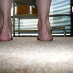
    Custom molded orthotics Part 4. So what really makes a custom molded orthotic made by your podiatrist foot and ankle expert different form the other outlets that are offering cheaper alternatives. First of all your podiatrist had four years of medical school training and on average two to three years of residency training on only foot and ankle. I myself did even an extra year of orthopedic training in limb lengthening and reconstruction. Now that we have the mold of your foot the next thing is to write the prescription to the laboratory on where the modifications need to be made. Look at the picture of this patients hind foot alignment, his heel everts (turns away from the leg)

    Pronated everted calcaneus
    Pronated everted calcaneus

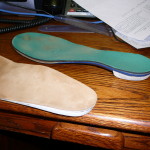
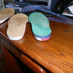
    the left foot is worse than the right, so the left foot orthotic prescription needs to have a different amount of correction of the pronatory forces. The doctor will tell the lab how much to adjust based upon his/her measurements. That’s not all does the person have a rigid stiff deformity or is it flexible. Patients tend to tolerate different orthotic materials based on if the foot is stiff and arthritic. The older one is usually they dont tolerate hard stiff orthotics. They work much better in children and patients up to thirty years old. Also modifications can be made to the arch height for flat feet , and the ball of the foot. Different degrees of tilt can unload different joints to give relief. If one is suffering from hallux limitus or hallux rigidus there are modifications that can be made such as kinetic wedges and cutouts to relieve the joint.

    Therefore when you are getting a custom device form a podiatrist you are getting a thorough exam of the foot and ankle and a prescription that is specific

    custom orthotic for posterior tibial tendon dysfunction
    custom orthotic for posterior tibial tendon dysfunction

    to address your deformity with careful consideration of age, activities and your personal needs. Custom molded orthotics can be a great way to get rid of your heel pain and plantar fasciitis.

  • Custom molded orthotics 3

    Custom Molded Orthotics 3

    What is so special about custom orthotics and what is done to them that makes them work? Why are they so expensive? These are all great questions that you should ask. The first step in making custom orthotics is the evaluation of your foot. Your Podiatrist will evaluate your foot for a plethora of positional deformities such as pronation, supination, forefoot valgus, forefoot varus, heel varus, and valgus. Also, the flexibility of your numerous joints is taken into consideration regarding orthotic evaluation. A standing exam and a walking gait exam are performed. Remember there are 33 joints in your foot and the flexibility or rigidity of your joints plays an important role. Once the doctor has an understanding of your foot type, he or she may decide an x-ray is important. For example, an x ray is the best way to tell if you have arthritis or a heel spur or plantar fasciitis, your local “expert” at the running store can not take an x-ray and interpret one. Also, an x ray will tell us if you have a stress fracture and the answer to your pain is not an orthotic but a cast or boot. Once a proper diagnosis is made then the patient’s foot shape is captured.

    There are 3 different ways to capture the shape of one’s foot. First is a plaster slipper cast mold ( today there are available fiberglass slipper socks that get great molds). The second method is a foam mold. I am not a big fan of the foam, because you lose accuracy in the shape of the foot. The 3rd and most accurate way is a 3-D scan of the foot. This is the preferred method at the Advanced Foot And Ankle Center Of San Diego. The reason is that it is the most accurate method and it gives us the most precise measurements Stay tuned for part 4 of this series.

  • MANOS Plantar Fascia Release System for heel pain

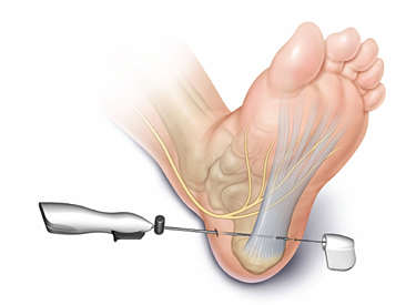
    Introducing the MANOS Plantar Fascia Release System for heel pain and plantar fasciitis. This is a new age for getting rid of heel pain that has not responded to injections and orthotics and night splints. This device when used releases the plantar fascia with a no stitches needed approach. Heel pain can be very frustrating  and sometimes you may spend hundreds of dollars on different devices such as orthotics and night splints. While those devices many times help there is a percentage of patients that fail those treatments. Most people are weary of surgery due to having large incisions and sutures and not being able to walk on their foot for weeks after. This new device called MANOS allows for the release of the plantar fascia with a no incision approach. Also you get to walk on it immediately after the procedure is performed. The patient is usually sedated so that you don’t feel anything and a local anesthesia is given as well. once the device is inserted the surgeon uses ultrasound to identify the plantar fascia. and the micro blade then releases the painful plantar fascia off of its calcaneal insertion. The patient needs no stitches and gets to walk on it right away. No more heel pain. We have had great success on many patients getting rid of their heel pain using minimally invasive procedures for heel pain and plantar fasciitis.

    MANOS Plantar Facia Release
    minimally invasive procedure for plantar fascitiis

    Not all the time will heal pain resolve with orthotics and steroid injections. Make sure that you are evaluated properly , I have had many a patient that has had tears of the plantar fascia and scarring that ultimately needed the band of tissue released. Feel free to contact me to see if you are a candidate. Heel pain no more.
  • Charcot Foot Reconstruction 1

    Charcot Foot Reconstruction 1

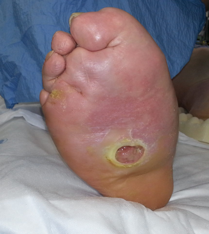
    Charcot foot reconstruction can be one of the most challenging surgeries of the foot and ankle. What is charcot foot ? How does one get it and what do you do if you end up with charcot foot?

    Charcot foot otherwise known as neuropathic arthropathy or neuropathic osteoarthropathy or charcot joint is a disease process that happens where the bones of ones foot or ankle start a process of resorption and soften which then leads to a breakdown of joints which then lead to deformity of the foot and ulceration’s. Not all feet ulcerate, however once the foot ulcerates the patient is at a high risk for infection and loss of foot or leg. This happens moslty in diabetics however it can happen in Alchoholic neuropathy spinal diseases certain steroid injections and syphillis.  the process by which this happens is a complex cellular reaction and microtruama which leads to the final charcot foot. Ultimately for today’s blog i would like to discuss  how we are

    Charcot foot
    Charcot foot

    treating these feet and preventing amputation. Usually these feet become deformed in the opposite direction of what a normal foot is. A normal foot has an arch to it however a charcot foot becomes a rockerbottom foot. Lets compare this to a suspension bridge, when looking at the road on the bridge from a distance the road looks like and arch but if the suspension is not holding up it looks sagging. Similar to what the foot bone structures look like when the charcot process completes itself.

    There are many ways to reconstruct a charcot deformity, in future blogs I will show you some of my cases and the use of the Taylor spatial frame particularly the Miter frame construct on how a two stage procedure is the safest method to obtaining the correction needed.

  • Plantar plate tear

    Plantar plate tear of the second metatarsal phalangeal joint is a condition of the foot that is often overlooked by foot doctors. Many people have come into my office that have been treated for neuroma’s and other ball of the foot pain that have been diagnosed incorrectly. Unfortunately people have spent hundreds of dollars on orthotics and injections and physical therapy when the issue was a Plantar plate tear.

    I will now go on to explain what the plantar plate of the second metatarsal phalangeal joint is and what it does. Then I will discuss the proper way to diagnose a tear of the plantar plate. After which i will discuss treatment options.

    What is the plantar plate.

    Above you see pictures of the second metatarsal phalangeal joint, that joint is a ball in socket joint that is surrounded by a capsule. Connected to the bottom portion of the capsule is a cushion material that the tendons slide under called the plantar plate. It is part of the padding on the bottom of the foot.

    This can become injured in many ways, I find it very prevalent in runners. People with certain foot types can be more susceptible to this condition. Such as if you have a long second metatarsal , which can only be diagnosed by x ray. Also people with flat feet and excessively high arched feet can get these tears.  Ultimately this can happen to anyone. Some times orthotics can prevent them or stop them from getting worse . As well minimalist shoes can make the problem worse as well due to the lack of support in that area.

    In the next blog in this series we will discuss how plantar plate tears are diagnosed and what are the treatment options. I am sure you are wondering is surgery necessary and are there ways to avoid surgery. Here at the Advanced Foot And Ankle Center Of San Diego we have thousands of patients that we have treated successfully with this condition. Feel free to contact me with any questions.

    platar plate of the 2nd MPJ
    plantar plate animation
  • Speeding up healing of Foot fractures and Ankle Fractures part 3

    Speeding up healing of Foot fractures and Ankle Fractures part 3

    Speeding up healing of foot fractures and ankle fractures are  the number one priority if this happens to you.

    Ankle Fracture
    Ankle Fracture

    A00391F06 In previous posts we discussed the three phases of bone healing and things that can slow down bone healing. In this post we will discuss methods to speed up bone healing if you sustain a fracture.

    There are devices known as bone stimulators. In essence what they do is stimulate the bone in such a way that they change the local biology and encourage fast healing . In my practice we have used them successfully in fresh foot and ankle fractures as well as non unions of fractures.

    There is the Exogen  and the Physio-stim and finally the CMF from DJ global.


    .

    The Exogen uses a low intensity pulsed ultrasound technology that upregulates Cox 2 , VEGF and BMP 2,4,6 and 7. It is the only

    exogen bone stimulator
    exogen bone stimulator

    This is an FDA approved bone stimulator for fresh (ones that just occurred) tibia and radius fractures, all the other ones are approved for non unions. Why would you want to wait till you get a non union to treat your fracture. Also it is applied for only 20 minutes a day. Patients love that. It has been shown to heal 86% of non unions and 81.6 percent of smokers with non unions that is important. Their studies show for foot metatarsal non unions a 92% healing success rate.

    Next is Physio-stim made by orthofix  which works by a pulsed electromagnetic field PEMF, their advantage is that it can go over a cast they show 85% overall healing through their published registry data on their website. The Pulsed electromagnetic field encourages new blood cell formation which helps bring the cells for the new bone growth. Their studies show for foot metatarsal non unions a 90.9% healing success rate. Ther device needs to be worn 3 hours a day.

    The CMF by DJ ortho  creates a low magnetic signal at the fracture site that stimulates new blood cells to bring new bone cells. It is to be worn for 30 minutes a day. No human studies demonstrate efficacy of CMF see  http://www.medscape.com/viewarticle/778366_7

    That is a review of the 3 major bone stimulators available today.