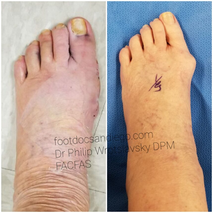
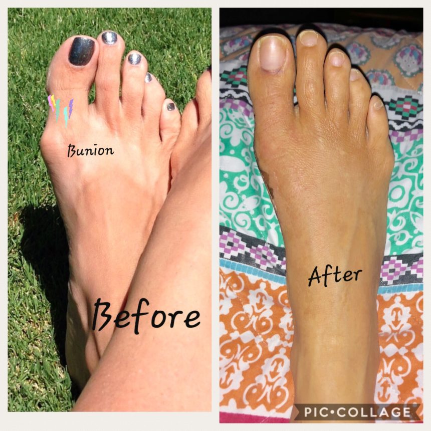
Are you considering bunion surgery? We just wanted to share with you some before and after pictures.
Bunion SurgeryBottom is before top is after this was done on both feetBefore and after bunionectomy
We use a technique that has no visible incision and has one of the fastest recovery times. Cosmetically pleasing results with no long term non weight bearing.
Bunion surgery ,bunionectomy, hallux valgus, bump on the big toe, hallux abducto valgus
Not all wounds of the foot need surgery. Too often doctors can be pre-programmed to tell a patient when faced with a difficult diabetic toe wound, to just amputate the toe. Amputation many times is the best solution, but not always.
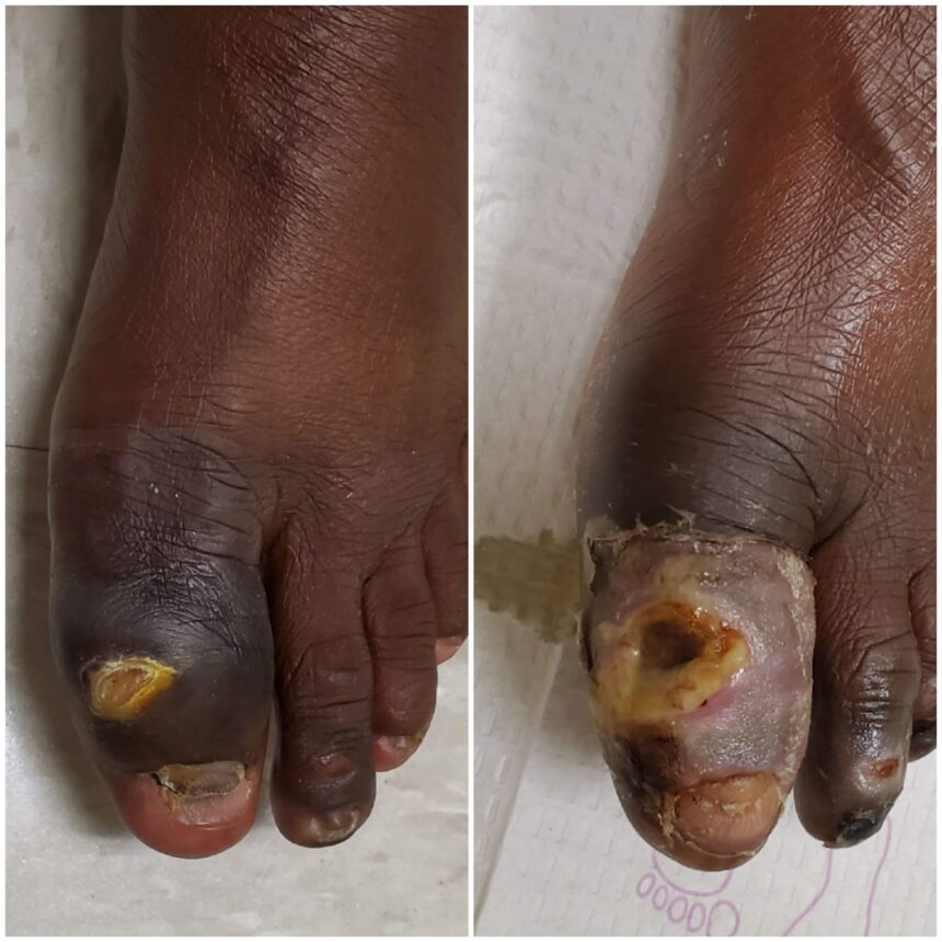
before and after diabetic toe wound
As can be seen in the picture above, the toe on the right is the initial presentation of the patient , the one on the left is a little then 2 months later.
The above patient presented with a deep penetrating diabetic toe wound and was at high risk for loss of a toe. X rays were positive for bone infection.
infected bone
Not all toes that are infected, even if the bone is infected, need amputation. This is a great case of no surgery needed. 6 Weeks IV antibiotics was all that was needed to get rid of the infection.
resolving infection
This case is a great example of not rushing to surgery, and utilizing a team approach of Vascular and Infectious disease specialists to prevent amputation.
I was recently asked to give a lecture on Charcot foot and ankle reconstruction and thought that this was an opportune time to write a blog about the new updates and treatments regarding Charcot foot and ankle reconstruction.
Charcot right foot and ankle reconstruction has undergone many types of treatment options over the past 100 years. We have moved far past needing to amputate patients that are affected with Charcot neuroarthropathy, as the first line of treatment. Most of the patients today can do well with reconstruction techniques that allow them to go back into her shoe or to be in a boot.
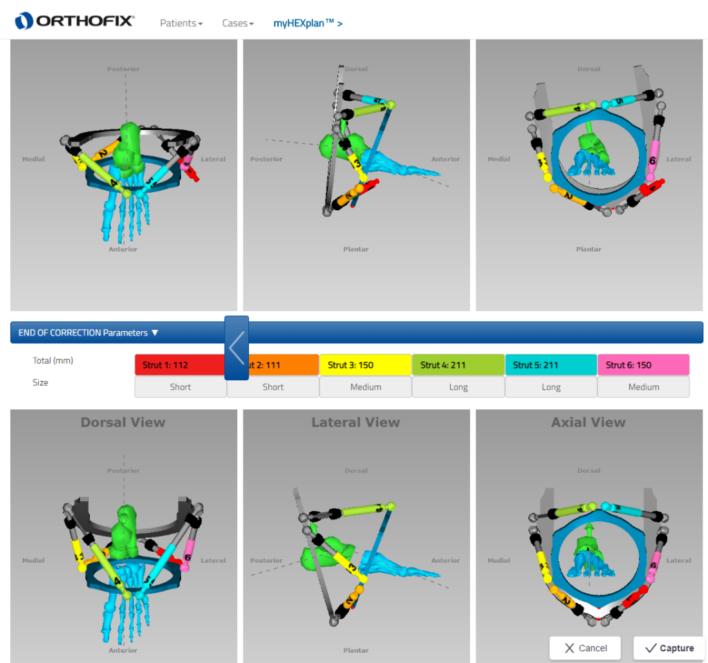
With regard to Charcot foot reconstruction, many advancements have been made in deformity analysis and deformity correction techniques that allow for minimal removal of bone and minimal removal of soft tissue that also allow for maintaining the length of the foot. We used various computer deformity programs that allow us to accurately measure how much bone needs to be removed as well as the precise area of where to make the bone cuts when necessary to reposition the bone after the collapse of a Charcot foot or ankle.
1 of the big advances that we have been able to make with Charcot reconstruction is the beaming technique. Intramedullary beaming of the foot and the ankle allow for a stable structure that makes it easier for the bones to heal. As opposed to in the past when big incisions were made with the application of plates with multiple screws the beams have better strength and from a mechanical standpoint hold a better than plates and screws. Due to the fact that people with Charcot have weakened bones, you will notice that many people Charcot have external fixators applied. Those external fixators are very important in allowing for the beaming procedures to be successful as it prevents 1 from moving the leg around or putting too much weight on their leg while the beams are working. Some people have very large deformities that require a technical gradual deformity correction. Gradual deformity correction is very similar to when 1 has braces for teeth. If one will go to an orthodontist on the orthodontist would say to them, that they were going to take your teeth and crank them into the right spot you get pretty nervous and run out of the office. What you may have expected to hear their input on braces that slowly take the teeth and put them back in the right position. Similar is with the foot and ankle there are many deformities that you cannot put back into the right position and 1 shot and requires a two-stage procedure. That is why you see some of these external fixators with rods next to them that the little rod used to turn very slowly and will slowly correct the foot in the right position. One of the big advancements that we have now with deformity correction are computerized programs that allow us to take exact measurements of where the bones are where the deformity is and we are exactly to make the bone cuts so that the healing is done in the most optimal fashion.
computer-assisted deformity correction.
Some of the updates that we find with ankle Charcot are similar to foot Charcot in the sense that the combination of rodding and external fixation is vital for recovery. This comes from the same idea that since the bones are very weakened with Charcot just putting in a plate with some screws is destined for failure. Most cases of ankle Charcot require an intramedullary rodding to hold it in place with an external fixator on top of that. Once the healing is complete and the external fixator can take it off and then you have the rod left and there. Most people with ankle Charcot end up doing better with this technique. Up until the last few years when this technique became more excepted many people had failures of ankle reconstruction secondary to Charcot surgery because this was not to the excepted technique. Now that this is the excepted technique we have gone from a 50% success rate to over 90% success rate.
Lastly, there are many other tricks and techniques that we have found that works very well with making sure the skin heals properly. There have been some great advancements and negative pressure therapy on incisions to help the incisions heal faster. Utilizing all of these techniques we were able to help people with Charcot foot and ankle get back to the regular activities. And at least prevent many amputations that are unnecessary because of these new techniques.
There are over 136 million people in North American that have diabetes or are pre-diabetic. If you are reading this, odds are you are one of them. When you have diabetes there are a lot of things going on that you must watch for. Many people with diabetes, about 70%, have a condition called neuropathy, which means you can’t feel your feet. When you can’t feel pain in your feet it means you can’t feel when there is an issue, like a rock in your shoe, laces tied too tight or too much pressure being placed on the bottoms of your feet. These situations can quickly develop into wounds, which if not managed appropriately can last for years and in the worse cases end up with an amputation. Orpyx orthotics can help prevent diabetic foot ulcers from recurring.
Somewhere in the world, a leg is lost to diabetes every 30 seconds, but the good news is 85% of the time this can be prevented with proper care. FINALLY, a solution!
Recently a new technology from Orpyx® Medical Technologies caught my attention. It is a sensory custom orthotic system that monitors movement, pressure, and temperature on the bottom of your feet. It provides real-time alerts and offloading instructions when sustained, high-pressure levels occur. This is all done through a mobile app that is downloaded to a smartphone or through a display device provided by the company. You and your healthcare provider can use this information to help prevent these wounds from recurring and keep your feet healthy. In fact, a study was recently published with people that had diabetes, neuropathy, and at least one wound in the past twelve months. During the 18 months of the study, these patients saw an 86% reduction in the diabetic foot ulcer reopening using the Orpyx® SI technology.
The best treatment plan for diabetic foot ulcers is to prevent them from happening. Orpyx SI sensory insole ( custom-molded orthotics) system is one way to help keep your feet healthy. Check them out! Orpyx custom-molded orthotics can help prevent diabetic foot ulcers from recurring.
When one presents with a diabetic foot infection there is always the fear of amputation. Diabetics that have infections in their feet are at significantly increased risk for amputation. The challenge is to make sure that the patient can be treated with his minimal amount of surgery as possible saving as much of the leg as possible.
Over the years research has been done in the field of predicting the severity of infection to see if there is a correlation with any markers in the blood or diagnostic studies that can tell us how bad the infection is. The studies can help us understand whether one needs surgery versus being able to treat with intravenous antibiotics. Usually, x-rays or an MRI or bone scan will tell us where the infection is to the extent of how far deep into the foot or up the leg the infection goes. There have been studies as to what markers in the blood he can tell us the severity of the infection. The first thing that we look at is one is white blood cell count as that is the primary marker to see if your body is fighting infection by producing white blood cells to fight the infection. There are other studies such as ESR and CRP that can tell us the severity of the infection.
Recently there has been published research into a marker in the blood called procalcitonin. Procalcitonin is the peptide precursor of the hormone calcitonin. It usually is elevated when there is an inflammatory stimulus from a bacterial infection. A recent study that was published in the Journal of diabetes research showed that increased procalcitonin levels at the time of admission to the hospital were a high indicator of amputation. This is a very important study for doctors and patients alike as when one presents with an infected diabetic foot amongst all the many tests that need to be performed procalcitonin levels are very useful in helping the doctor and the patient be able to predict the proper treatment.
foot wound
This new data helps us create a better treatment plan with diabetics that have infections in the foot. Instead of waiting around for surgery or debridements, when this data is available more definitive treatment can be offered to the patient and prevent amputations. No one ever wants an amputation but given the choice of amputating a toe versus the whole leg, it seems obvious that one would rather lose the toe. With the information available from the procalcitonin test that can be the difference between losing the toe or leg.
Ankle pain can really hold you back. If one takes three to five thousand steps a day that’s 3-5000 times that your ankle can hurt you daily. That is a painful way to live. There are many reasons for ankle pain. One that we specialize in here at the Advanced Foot And Ankle Center Of San Diego is Osteochondral Defect of the Talus.
https://youtu.be/qRl2EmMhRmQ
As you can see from the video above this is a treatable condition. However one must realize that not many doctors are trained in and not many doctors have much experience in doing these procedures.
The first challenge is getting the diagnosis. Once one is evaluated in the office an x-ray is helpful in pointing us in the right direction. Many times the x-ray is insufficient, the next step is an MRI. The MRI is more specific for cartilage disorders. As can be seen below the dome of the talus has lost its natural contour and there is a defect of the cartilage of the ankle joint.
Osteochondral Defect of the Talus MRI
Once a diagnosis is made, then based on the size and position of the defect as well as pain levels associated with the defect then one can make an informed decision with their surgeon as to the proper treatment. This article is only about surgery because the defect does not repair itself and does not self-heal. Either you have pain or you don’t.
The technique I would like to share with you is performing a medial malleolar osteotomy to access the talus and properly repair the defect. In my opinion, this is the only way to fully visualize the talus and repair the cartilage. Using arthroscopy or any small camera will not reach far back into the joint to visualize is well enough. And if you can not see what you are repairing then you can not repair it.
We use special cryopreserved cartilage that is called cartiform. This implant has been highly successful in my hands in repairing cartilage injuries of the foot and ankle.
Medial malleolar osteotomy for Osteochondral defect of the talus
Above you can see in the picture the injured cartilage that is lifted off the bone.
Repaired Osteochondral defect of the talus with cartilage graft
Above you will see the cartiform placed over the area of the talus that had the cartilage injury.
Ankle injuries can be very serious and should not be ignored. Come in today for your evaluation.
Charcot foot requires expert treatment from surgeons that have experience treating this condition. When one is diagnosed with Charcot foot they should seek out a doctor that can show them how many of these they treated successfully. One with Charcot foot or ankle is at a high risk for loss of limb due to many factors associated with the disease. We have developed techniques that allow for accurate and reproducible treatments to save a limb diagnosed with charcot foot.
When one is told as part of their Charcot foot treatment that they need to live in a boot (sometimes known as a CROW boot), this is helpful to prevent wounds. However this treatment does not correct the deformity associated with the disease. Unless one corrects the deformity one is always at risk to develop the ulcer and lose the leg.
There have been many advancements in treating charcot foot deformities. Dr Wrotslavsky lectures nationally and internationally regarding proper treatment and techniques . We are able to use computer models to analyze the deformity and slowly correct the deformity. This puts much less stress on the nerves and arteries and veins in the area. Through these and other methods we can put one in a much better position to prevent amputation in charcot foot.
If you have a foot wound and it is taking too long to heal , come in for another opinion today. Many of these wounds have other underlying issues that once addressed will get rid of the wound .
Sprained ankle, how about if you sprain your ankle multiple times a year. Are you one of the millions of people that suffer with multiple ankle sprains and chronic ankle instability. Do you wonder why whenever you are about to go out on a hike you are afraid to roll the ankle. Many ankle sprains and other ankle injuries can get better on their own or with physical therapy. There are times you will need a brace to stabilize the ankle. Unfortunately there are people out there that have not gotten relief from those methods and continue to roll their ankle. I have developed advances in surgical stabilization of the ankle via a minimal incision approach. This allows for the patient to recover faster and easier.
It is vital for a patient to have the proper workup to evaluate the extent of the ligament damage. Ankle Range of motion, inversion , eversion , anterior draw sign are important parts of the physical exam. Radio graphic exams should include the typical 3 views of the ankle but should also include a stress inversion view. The stress inversion view can be better than an MRI. As it can tell you if the ligament is functioning. Just because an MRI says the ligament is intact it does not mean that it works. In a stress inversion view the tibio talar joint should not make an angle of greater the 5-7 degrees.
Ankle stress inversion view
Here at the Advanced Foot And Ankle Center Of San Diego we have developed surgical techniques that fix the issue using grafts that stabilize the ankle and allows for full function. Most importantly we use minimally invasive approaches that allows for quicker recovery.
Ankle Stabilization surgery
If you are suffering with multiple ankle sprains or ankle instability , make an appointment today. We treat anyone from professional athletes to your weekend warrior.
Gout attack in the big toe. That is what no one wants to feel. It can get so painful that even when your bed sheets touch your foot the pain is unbearable. Do not ignore the symptoms of big toe pain. If left alone one can end up with severe joint damage . There are many treatments available for the pain. From oral medications to injections. Today with medicines such as krystexxa and sprix one can control the gout symptoms and pain.There are times when surgery is necessary . That can happen when the gout develops into joint arthritis and the to can not move anymore. The disease process is a buildup of crystals do to excessive uric acid build up. Through diet and watching lifestyle habits one can prevent the flare ups. However there are times that the buildup of the crystals or gouty tophi get to be so great that surgery is necessary to remove them. An X ray can be very helpful along with blood work to evaluate the extent of the gout. Many times as in the picture above one can see there looks like a growth on the foot. That is a buildup of the gouty tophi crystals. I have seen patients that the crystals pop out and start draining and a big wound is created on the foot. Do not hesitate to come in if you have big toe pain.
Bunions are not pleasant to say the least. First you have the painful bump that bothers you after a day of walking around. Then there is the issue of finding proper fitting shoes. Lastly there are the mechanical issues including a stiff joint and over lapping or under lapping of the second toe. There are many treatment options available. Non surgical ones include wider shoes and anti inflammatory medications. There are times when one gets so frustrated with their bunions that surgery becomes necessary. Here at the Advanced Foot And Ankle Center of San Diego we use techniques that make the recovery very easy. Many procedures today allow for immediate weight bearing. So no more ” you need to be off your feet for 12 weeks”. Also we offer minimal incision approaches that there is virtually no visible scar. As you can see from the picture on the right there is no visible incision, the big toe is not riding on the second toe and lastly that pesky bump is gone. Using our advanced techniques we are able to minimize post operative pain, this is very exciting for patients that have had painful surgeries in the past. I have had many patients come in that have had bunion surgery elsewhere and wanted to have the other foot done at our center, time after time they have told me that the pain was much less at our institution.
Hallux Varus
Very often I will have people that come in that have failed bunion surgeries from other institutions. The above picture is a from another surgeon that had a complication with her bunion surgery. The surgeon over corrected the bunion and created the varus deformity that you see in the picture. We experts in performing revisions of non unions and Hallux Varus complications. Many of my patients come to me with failed foot surgeries from elsewhere that need revision surgery.
We also have 3-D bunion correction available. With the Lapiplasty procedure we are able to get the most accurate correction possible. This is a big breakthrough in bunion surgery.
https://vimeo.com/199272477
If you have a bunion and have been told that surgery will entail too long of a recovery or is too painful come in for an evaluation today.